Acquisition guidance for facial CT or CBCT studies used in virtual surgical planning and related 3D workflows.
01
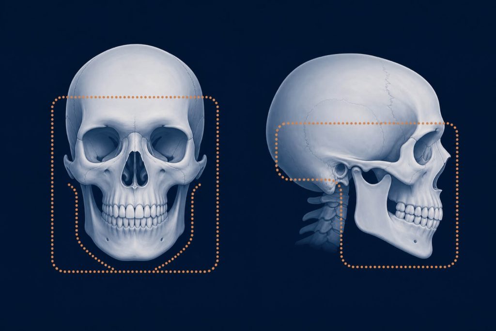
Anatomical coverage
Capture the complete planning anatomy.
The scan should include the orbits, glabella, mastoids, both temporomandibular joints, maxilla, and mandible.
Copper boundary: include the complete planning anatomy in both frontal and lateral extent.
02
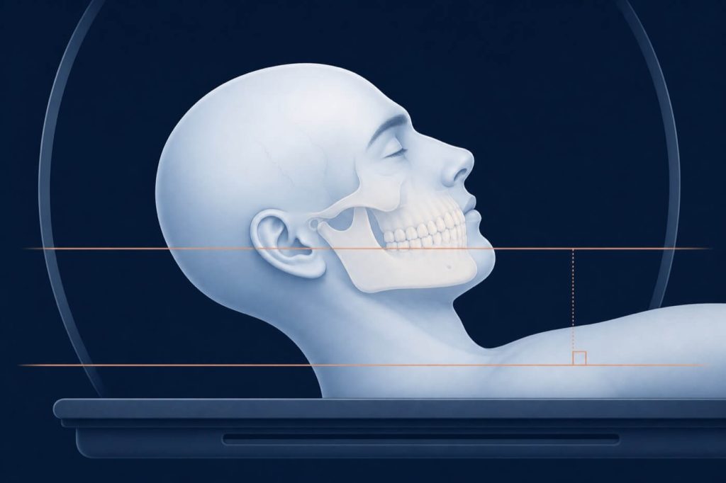
Patient position
Normal occlusion. Neutral alignment.
Copper guides: keep the occlusal plane parallel to the gantry or table reference.
Scan in normal occlusion, or use a thin wax wafer while maintaining normal occlusion.
Keep the occlusal plane parallel to the gantry to limit metallic restoration artefact around the teeth.
For CBCT, avoid chin cups or straps that deform the soft tissue.
03
Before acquisition
Essential scan checklist
Acquire at high spatial resolution with thin, contiguous slices of 1 mm or less.
Capture the original scanning plane at 0° gantry tilt.
Include the orbits, glabella, mastoids, both TMJs, maxilla, and mandible.
Use the largest available field of view for CBCT while including the complete anatomy of interest.
Keep the patient completely still; restart the scan if motion artefact occurs.
Remove metal items and jewellery from the head and neck region.
Provide the complete study as uncompressed, original DICOM data.
Use a scan that is less than three months old.
04
Technical reference
Recommended scan parameters
Parameter
CBCT
CT Scan
Matrix
512 × 512
512 × 512
Resolution
Highest available
Spacing < 1 mm
Voxel / Pixel
0.3–0.5 mm voxel
Pixel < 0.6 mm
Field / Tilt
Largest FOV available
0° gantry tilt
Series
Axial reconstruction
Original / primary / axial; no reformatting or post-processing
CBCT
CT Scan
Matrix
512 × 512
512 × 512
Resolution
Highest available
Spacing < 1 mm
Voxel / Pixel
0.3–0.5 mm voxel
Pixel < 0.6 mm
Field / Tilt
Largest FOV available
0° gantry tilt
Series
Axial reconstruction
Original / primary / axial; no reformatting or post-processing
Important
Send the complete, uncompressed DICOM study in its original format. Screenshots, compressed image exports, or reformatted series are not suitable substitutes for planning data.
A consistent photo set for virtual surgical planning, alignment, and facial-dental reference.
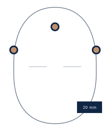
Required scale reference
Place a 20 mm marker at three points.
Every photograph must show a 20 mm marker on the forehead and on both temple regions. Keep all three markers clearly visible in every required photograph.
01
3D CT / CBCT DICOM
Face straight, with the patient’s eyes directed consistently for the scan.
02
Dental cast STL
Provide scans of the existing occlusion and the desired occlusion.
03
Frontal · repose
Head straight, eyes looking ahead, and lips in repose.
04
Frontal · smile
Head straight with the patient’s natural smile.
05
Lateral · repose
Capture both left and right lateral views with lips in repose.
06
Lateral · smile
Capture both left and right lateral views with a natural smile.
07
Intraoral views
Document the patient’s existing occlusion with clear intraoral photographs.
─────
Need Help Before Acquisition?
Confirm the protocol before the patient arrives.
If the clinical situation or modality differs from the standard workflow, ask the EDIS planning team to confirm the required records.
Case-specific guidance
Confirm variations with the EDIS planning team before acquisition.
────
Submit a case
Tell us what
the case needs.
We will review the details and respond within one working day with next steps.
• 12,000+ Cases Delivered • Fast Turnaround • Clinician Reviewed
No payment is required. We will confirm scope, timing, and cost before work begins.